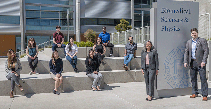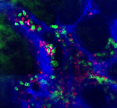
Immunology Professor Jennifer Manilay and bioengineering Professor Joel Spencer are using a new grant from the National Institutes of Health (NIH) to expand a project they’ve been working on for the past two years — delving into the immune systems of living mice to see how B-cells develop under different circumstances.
B-cells are a type of white blood cell that are critical to the immune system because they determine the specificity of the immune response to infectious microorganisms and other foreign substances.
Thanks to Spencer’s cutting-edge imaging technique that allows the researchers to see inside a living mouse’s bone marrow, the cross-disciplinary team can put several ideas to the test.
“We know that osteocytes contribute to B-cell development, but we hypothesize that mesenchymal stem cells and pre-osteoblast cells are also critical,” Manilay said. “Mesenchymal cells develop into pre-osteoblasts, and the pre-osteoblasts develop into osteocytes, so we want to look at each stage of development.”
To do so, the team will obtain mouse sperm from a German collaborator to breed mice that meet certain genetic specifications and will also work with UC Davis to “build” a reproductive pair of mice that will allow investigation of B-cell development using an inducible, conditional gene knockout approach. That will allow the researchers to introduce a certain enzyme that will delete, or knock out, the VHL gene in specific cells at specific times.
The researchers will breed the mice and examine the bone marrow systems of the pups at three-week intervals to see how the B-cell development is affected when the VHL gene in mesenchymal cells and pre-osteoblasts is deleted.
“There’s a lot of technology going into this project,” Manilay said.

It’s important to be able to control the timing and spacing of the gene knockouts so the researchers can look at the same mice over time, and test for different situations.
“VHL helps control cells’ response to different oxygen levels, especially low levels. Our current data indicates that VHL gene deletion accelerates the production of red blood cells and helps create blood vessels in the bone and bone marrow,” Spencer said. “This, in turn, impacts how B-cells develop.”
The researchers will be able to peer inside the bone marrow and see how the microenvironment has changed when VHL is deleted in the mesenchymal cells and pre-osteoblasts. Several diseases are linked to the marrow microenvironment, so the research has the potential to inform the work of many other research teams — as could the development of the new mouse model. Manilay said many researchers have tried to build models with the same specifications she and Spencer need, but those models have not worked.
“We’re trying something different, so this could really help researchers all over the world if it works,” she said. “This is high-risk, high-reward research. The risk is this could completely fail, but even then, we’d be able to provide information to other researchers.”
This $400,000, two-year R21 grant builds on an NIH R15 award the team received last year to examine the effects of the VHL gene defect that makes the bones grow extremely dense, with little room for marrow. Because marrow is where both immune system and blood cells develop, less marrow than normal could have a wide range of negative consequences, and the team wanted to know whether immune cells were responding to altered oxygen conditions, which are also a hallmark of the defect. B-cells can sense when they are in low-oxygen conditions, Spencer said, and they can turn on genes to help them adapt.
Spencer, whose research focuses on biomedical imaging, became the first scientist to capture an image of native adult hematopoietic stem cells (HSC) within the bone marrow of a living organism. Manilay is interested in the relationship between bone and bone marrow’s HSCs on immune cells’ fates.
Spencer, with the Department of Bioengineering in the School of Engineering, and Manilay, with the Department of Molecular and Cell Biology in the School of Natural Sciences, are both members of the Health Sciences Research Institute. They worked on the foundation of this research for more than a year before the first grant started. Manilay’s lab had gathered preliminary data, including some work done by graduate student Betsabel Chicana, who recently won an NIH fellowship for her immunology work. The graduate students collaborated throughout, analyzing data and cross-training in each other’s disciplines.
“Now that we’ve been doing this for more than a year, I’m really seeing the benefits among my students,” Spencer said. “Exposing them to other research methods and other ways to ask questions and design experiments gives them a broader training, which will benefit their futures as researchers.”
Lorena Anderson

Senior Writer and Public Information Representative
Office: (209) 228-4406
Mobile: (209) 201-6255


